Volume: 14 Issue: 2
Hydatid Cysts at Uncommon Anatomical Sites: A Histopathological Case Series from Central India
Year: 2026, Page: 103-106, Doi: https://doi.org/10.47799/pimr.1402.26.38
Received: March 28, 2026 Accepted: June 29, 2026 Published: Aug. 31, 2026
Abstract
Hydatid disease caused by Echinococcus granulosus most commonly involves the liver and lungs; however, primary involvement of atypical anatomical sites is uncommon and often poses diagnostic challenges. This retrospective case series describes four patients diagnosed with hydatid cysts at rare locations, including the orbit, gluteal muscle, kidney, and pancreas, at a tertiary care center in Central India. All cases presented as cystic masses clinically mimicking other benign or neoplastic conditions. Definitive diagnosis was established on histopathological examination demonstrating characteristic laminated cyst walls, germinal membranes, and protoscolices or hooklets. Serological investigations were not uniformly available, which is acknowledged as a limitation. This series highlights the importance of considering hydatid disease in the differential diagnosis of cystic lesions at unusual sites, particularly in endemic regions.
Keywords: Hydatid cyst; Echinococcus granulosus; Unusual anatomical sites; Histopathology; Zoonotic infection
INTRODUCTION
Hydatid cysts, a zoonotic parasitic infection, remains a significant global public health concern. Recognized by the World Health Organization (WHO) as a neglected tropical disease, it predominantly affects regions such as South America, East Africa, Central Asia, China, and the Mediterranean[1]. Human incidence rates can reach up to 50 per 100,000 person-years, with prevalence as high as 5–10% in certain communities[1]. Common intermediate hosts are sheep, cattle, and pigs. Humans may accidentally become hosts by ingesting parasitic eggs containing the larval stage of the tapeworm Echinococcus granulosus. While the cysts typically affect the liver (50-77%), the other organs involved are the lungs (15-47%) and spleen (0.5 - 8%)[1]. Occasionally, hydatid cysts occur at rare locations[2]. The clinical presentation of hydatid cysts varies depending on the organ involved and may lead to significant complications and morbidity. This case series highlights four cases of primary hydatid cysts in atypical locations, emphasizing the need for clinicians to consider this diagnosis when encountering cystic lesions at uncommon sites.
MATERIAL AND METHODS
This retrospective case series included patients diagnosed between 2021 to 2024 whose surgical specimens were received in the Department of Pathology with a final diagnosis of hydatid cyst at uncommon anatomical sites. Clinical presentation, imaging findings, operative details, and available follow-up data were retrieved from medical records.
Serological investigations for hydatid disease, including anti-Echinococcus IgG or IgG4 assays and antigen detection tests, were not consistently performed, as the cases were referred primarily for histopathological evaluation following excision. Diagnosis in all cases was confirmed by characteristic microscopic findings. Given the retrospective and surgically referred nature of these cases, histopathology was considered the diagnostic gold standard.
Case Series:
All four patients presented with localized symptoms related to the anatomical site involved. None had a prior clinical diagnosis of hydatid disease at presentation. From a clinical standpoint, the provisional differential diagnoses included benign cysts, chronic inflammatory lesions, abscesses, and site-specific neoplastic conditions. Hydatid disease was not suspected preoperatively in any case due to the unusual locations.
Case 1: An Orbital Hydatid Cyst A 4-year-old male presented with a mass in the left upper eyelid. Clinically and radiologically, the lesion was suspected to be a benign cystic mass. Lesion was measuring 2.2 × 2.2 × 1 cm, with a mucosal covering of 1.2 × 0.5 cm. Gross examination revealed a partially cystic mass filled with necrotic tissue. Microscopic analysis revealed a laminated cyst wall with a germinal epithelial lining surrounded by dense inflammatory infiltrates and foreign body giant cells.
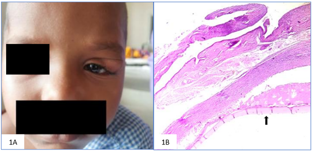
Fig. 1: A. Case 1: Orbital hydatid cyst showing cystic mass. B. Case 1: Cyst wall with laminated membrane (arrow) and foreign body giant cell reaction (H&E, 10 X)
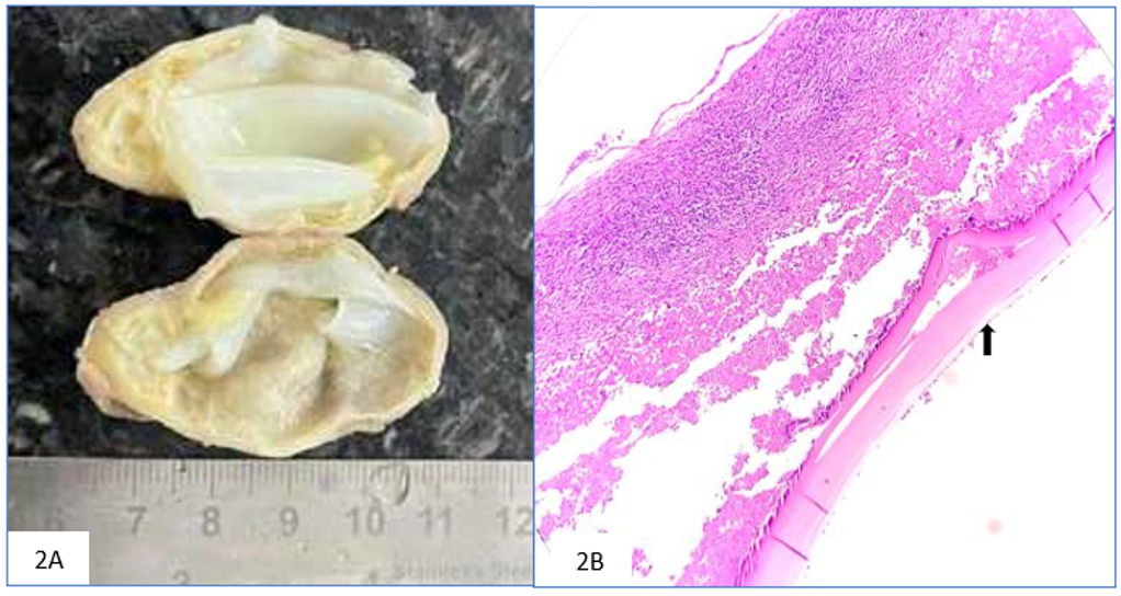
Fig. 2: A. Case 2: Gluteal hydatid cyst with clear fluid-filled cavity. B. Case 2: Laminated cyst wall with germinal layer (arrow) and inflammation (H&E, 10 X)
Case 2: Gluteal Hydatid Cyst A 33-year-old female presented with a left gluteal nodular swelling. Clinically suspected to be cystic lesion. The excised mass measured 5 × 3 × 2 cm and contained clear, watery fluid. Microscopic examination revealed a laminated cyst wall membrane, with a germinal membrane surrounded by dense inflammatory infiltrates.
Case 3: Renal Hydatid Cyst A 33-year-old female was diagnosed with a cyst in the left kidney. Clinically and radiologically, the lesion was suspected to be a benign cystic renal mass. On gross examination the cyst, located at the lower pole, measured 8 × 7 × 5 cm, with an overall kidney size of 11.5 × 8 x 4.5 cm. Microscopic analysis revealed a hyaline eosinophilic acellular lamellate layer containing scolices and hooklets. The wall of the host cyst showed a desmoplastic reaction.
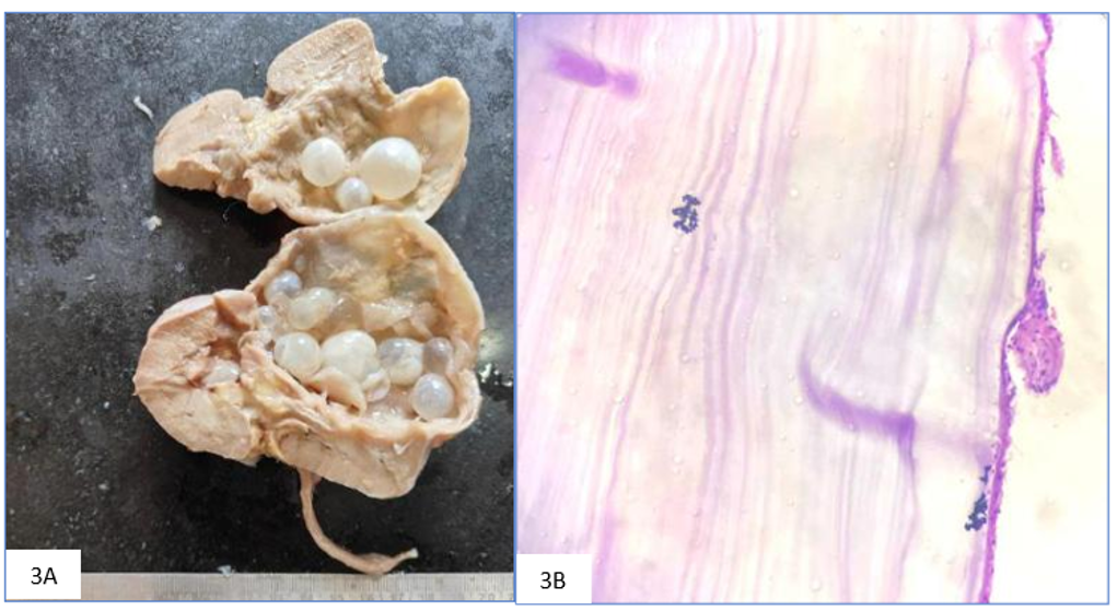
Fig. 3: A. Case 3: Renal hydatid cyst involving lower pole of kidney. B. Case 3: Cyst wall showing scolices and hooklets with desmoplastic reaction (H&E, 40 X)
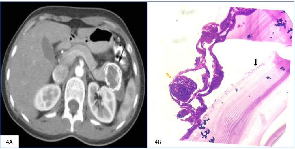
Fig. 4: A. Case 4: Pancreatic hydatid cyst with nodular cystic lesions (CT Scan, Abdomen). B. Case 4: Cyst wall with laminated membrane (black arrow), germinal lining, and protoscolices (yellow arrow)
Case 4: Pancreatic Hydatid Cyst A 24-year-old female presented with a cystic mass in the tail of the pancreas, measuring 7.5 × 7 × 6 cm. Clinically and radiologically, the lesion was suspected to be a pancreatic cystic neoplasm. Gross examination revealed the presence of multiple nodular cystic lesions. Microscopic analysis revealed a laminated cyst wall membrane with a germinal membrane and protoscolices accompanied by inflammatory reactions in solid areas.
All patients underwent complete surgical excision; perioperative antihelminthic therapy was administered where clinically indicated. Information regarding epidemiological risk factors such as direct contact with dogs or livestock was not uniformly documented in the medical records. Postoperative follow-up details were limited due to the retrospective nature of the study.
DISCUSSION
Hydatid cysts are a significant health concern in many regions, with the liver and lungs being the most commonly affected organs. However, this case series highlights the potential for hydatid cysts to develop at unusual locations. The incidence of atypical sites is estimated to be 8-10% in all hydatid cases[3]. Orbital hydatid cysts account for 0.7% to 1% of cases[4], whereas pancreatic hydatid cysts are rare, with an incidence ranging from 0.14% to 2%[5]. Renal involvement is observed in 1–3% of hydatid cases[5]. Cysts are typically located at the upper or lower poles and are usually unilateral. Muscle involvement occurs in 0.5% to 4% of cases[6].
These unusual presentations pose diagnostic challenges as they may mimic other cystic lesions or tumors. A high index of suspicion is crucial for an accurate diagnosis, particularly in endemic areas. Imaging, serology, and histopathological examinations play vital roles in confirming the diagnosis.
Serological tests such as ELISA for anti-Echinococcus IgG, IgG4 subclass detection, and circulating antigen assays are recommended adjuncts in the diagnosis of hydatid disease. However, their sensitivity varies depending on cyst location, stage, and host immune response. In cysts located at atypical sites, serological tests may yield false-negative results due to low antigen burden or sequestration of cyst contents[7-8].
Several studies have reported hydatid cysts in uncommon anatomical locations, highlighting the varied presentations of this parasitic cyst. In a recent case reported by Yalavarthi et al. in 2013[6], hydatid cyst presented as an intramuscular cyst in the thigh region, which is a rare but well documented. Kothiya et al. in 2022[2] described pancreatic involvement. Similarly, Arora, Sarngal, and colleagues in 2022[9] observed hydatid cysts in diverse sites including the axillary subcutaneous region, ovary, gallbladder, and pancreas, indicating that the cyst can mimic a wide range of pathological conditions. In 2021, Ma et al.[4] reported the involvement of the orbit and central nervous system (CNS), both of which are highly atypical sites and can pose significant diagnostic challenges. In 2014, Sachar et al.3 documented hydatid cysts involving the left hypochondrium, kidney, mesentery, and right inguinal region, whereas Attash in 2013[10] noted cases involving the pancreas, abdominal wall, spleen, back, and thigh. These reports collectively underscore the capacity of Echinococcus granulosus to affect almost any organ system and emphasize the need for heightened clinical suspicion and diagnosis, especially in endemic areas.
All cases originated from a region endemic for hydatid disease; however, the institutional base rate of hydatid infection could not be calculated due to the absence of centralized epidemiological data. No specific patient-related risk factors could be identified that explained the unusual anatomical distribution, consistent with previous reports suggesting hematogenous dissemination as a possible mechanism.
Limitations of the study:
The limitations of this study include its retrospective design, incomplete serological evaluation, limited clinical follow-up, and reliance on histopathology-based case identification. These factors restricted detailed analysis of clinical management and outcomes.
CONCLUSION
This case series demonstrates that Echinococcus granulosus can affect virtually any organ in the body, with a few exceptions such as hair and nails. The varied presentations of hydatid cysts in unusual locations underscores the importance of their inclusion in the differential diagnosis of cystic masses, regardless of their anatomical site. Healthcare professionals should maintain a high index of suspicion, particularly in endemic regions, because early diagnosis and appropriate management are essential to reduce the morbidity associated with this parasitic infection.
DISCLOSURE
Declaration of Patient Consent
Written informed consent was obtained from all patients and/or their legal guardians for publication of clinical data and images.
Data Availability: Collected data are available at the Department of Pathology, Raipur Institute of Medical Sciences, Raipur, Chhattisgarh, India.
Authors’ Contributions
Dr. Chandni Krishnani conceptualized and designed the study, drafted the initial manuscript, performed the literature search, reviewed patient data, and provided diagnostic expertise including histopathological interpretation and photomicrographs. Dr. Anil Kumar Verma and Dr. Firoz Sheikh critically reviewed and revised the manuscript for important intellectual content. Dr. Abhilasha Wahne performed procedures, collected clinical and radiological details, and assisted in manuscript preparation. Dr. Prateeti Pandey participated in patient care, compiled clinical details, and contributed to the literature review. All authors reviewed and approved the final manuscript and agreed to be accountable for all aspects of the work.
References
1. World Health Organization. Echinococcosis [Internet]. Geneva: World Health Organization; 2023 [cited 2025 Aug 1]. Available from: https://www.who.int/news-room/fact-sheets/detail/echinococcosis
2. Kothiya PK, Gupta V, Sarawagi R, Jayashankar E, Sharma J, Wani H, et al. Isolated primary hydatid cyst of the pancreas: Management challenges of a cystic masquerade. Annals of Hepato-Biliary-Pancreatic Surgery. 2022; 26 (4). Available from: https://doi.org/10.14701/ahbps.22-031
3. Sachar S, Goyal S, Goyal S, Sangwan S. Uncommon locations and presentations of hydatid cyst. Annals of Medical and Health Sciences Research. 2014; 4 (3). Available from: https://doi.org/10.4103/2141-9248.133476
4. Ma MS, Su F, Hei Y, Feng MY, Yan XJ. Disseminated hydatid disease in the orbit and central nervous system. International Journal of Ophthalmology. 2021; 14 (9). Available from: https://doi.org/10.18240/ijo.2021.09.25
5. Kothiya PK, Gupta V, Sarawagi R, Jayashankar E, Sharma J, Wani H, et al. Isolated primary hydatid cyst of the pancreas: Management challenges of a cystic masquerade. Annals of Hepato-Biliary-Pancreatic Surgery. 2022; 26 (4). Available from: https://doi.org/10.14701/ahbps.22-031
6. Yalavarthi S, Satya N, Ramamurti T, Supriya M. Intermuscular hydatid cyst in the thigh: An unusual presentation. Medical Journal of Dr. D.Y. Patil University. 2013; 6 (2). Available from: https://doi.org/10.4103/0975-2870.110311
7. Sarkari B, Rezaei Z. Immunodiagnosis of human hydatid disease: Where do we stand?. World Journal of Methodology. 2015; 5 (4). Available from: https://doi.org/10.5662/wjm.v5.i4.185
8. Zhang W, McManus DP. Recent advances in the immunology and diagnosis of echinococcosis. FEMS Immunology & Medical Microbiology. 2006; 47 (1). Available from: https://doi.org/10.1111/j.1574-695x.2006.00060.x
9. Arora S, Sarngal S, Gandhi S, Sharma S. Unusual Presentation of Hydatid Cyst. Euroasian Journal of Hepato-Gastroenterology. 2022; 12 (1). Available from: https://doi.org/10.5005/jp-journals-10018-1369
10. Attash SM. Unusual sites for primary hydatid cysts: self experience with five cases. BMJ Case Reports. 2014; 2014 Available from: https://doi.org/10.1136/bcr-2013-201843
Copyright
©2026 Krishnani et al. This is an open-access journal, and articles are distributed under the terms of the Creative Commons Attribution License CC-BY 4.0. (https://creativecommons.org/licenses/by/4.0/) which permits unrestricted use, distribution, and reproduction in any medium, provided the original authors and source are credited.
Cite this article
Krishnani C, Verma AK, Sheikh F, Wahne A, Pandey P. Hydatid Cysts at Uncommon Anatomical Sites: A Histopathological Case Series from Central India. Perspectives in Medical Research. 2026; 14(2):103-106 DOI: 10.47799/pimr.1402.26.38