Volume: 14 Issue: 2
Effect of Scapulothoracic Exercises in the Treatment of Adhesive Capsulitis: A Randomized Controlled Trial
Year: 2026, Page: 85-91, Doi: https://doi.org/10.47799/pimr.1402.26.118
Received: Aug. 6, 2026 Accepted: Aug. 27, 2026 Published: Aug. 31, 2026
Abstract
Introduction: Adhesive capsulitis is a chronic condition marked by inflammation and stiffness of the shoulder joint. This leads to a significant loss of both active and passive movement. While most rehabilitation approaches focus on the glenohumeral joint, this study also evaluates the role of the scapulothoracic region in shoulder mobility. Objectives: This study evaluates the effectiveness of adding targeted scapulothoracic exercises to conventional occupational therapy. Methods: 16 participants were divided into a control group receiving standard therapy and an experimental group receiving additional scapulothoracic exercises. Outcomes were assessed at baseline, 3 weeks, and 6 weeks, with pain scores, range of motion, muscle strength, and quality of life being the outcome measures. Results: Both groups showed significant improvements (p < 0.01); however, the experimental group demonstrated better results in several quality-of-life domains, including physical functioning, energy levels, social functioning, pain, and general health. Conclusion: These findings suggest that incorporating scapulothoracic exercises can enhance overall recovery and improve patient well-being in adhesive capsulitis.
Keywords: Adhesive Capsulitis, Frozen Shoulder, Occupational Therapy, Physical Rehabilitation, Quality of Life, Scapulothoracic Rhythm
INTRODUCTION
Adhesive capsulitis is characterised by pain and progressive decrease of both active and passive range of motion (ROM) of the shoulder joint. There is capsular thickening, fibrosis, and the formation of adhesions, leading to significant limitation of joint mobility. There are no arthritic changes in the cartilage or bone. The commonly affected movements are external rotation and abduction of the shoulder[1, 2]. Some of the symptoms reported are pain during movement and at rest, nocturnal pain, disturbed sleep, and functional limitations affecting activities of daily living (ADL). As there is a loss of glenohumeral joint motion, patients often make compensatory postural changes, such as scapular protraction, anterior tilt, rounded shoulders, and elevation. We may also see generalised muscle weakness, reduced endurance of the glenohumeral musculature. The position and movement of the scapula on the thorax are important for normal glenohumeral function and help in optimal shoulder movements[3-6]. The scapula provides a stable base for glenohumeral movement. Scapular stabilisers, including the Levator Scapulae, Rhomboids major and minor, Serratus anterior, and Trapezius, help in positioning the scapula so that efficient glenohumeral movement occurs. These muscle groups work in synergy with the rotator cuff to control scapular movement[3, 8]. When weakness or dysfunction is seen in the scapular musculature, normal scapular positioning and mechanics are disturbed, resulting in abnormal stresses to the capsular structures, rotator cuff compression and reduced performance[3, 5]. We have seen that new concepts in shoulder rehabilitation consider dynamic stabilisation of the scapula as a vital aspect of clinical management[3, 9]. Depression, downward rotation, and posterior tilt of the scapula decrease with age. There is a restriction of the scapular motions of depression, downward rotation, external rotation, and posterior tilt in adhesive capsulitis[10, 11]. Compensatory scapulothoracic movement increases as glenohumeral movement decreases[12]. There is a decrease in scapular stabilisation and an increase in the external rotation and protraction of the scapula[3, 4]. Such kinematic alterations can affect normal scapulothoracic rhythm. Conventional Occupational Therapy intervention programs primarily focus on improving glenohumeral joint ROM and muscle strength through functional activities[13, 14]. However, these approaches may not adequately address pain and dysfunction arising from altered scapulothoracic mechanics and compensatory muscle overuse. Although previous studies have explored the effects of scapulothoracic exercise programs on shoulder ROM in adhesive capsulitis, limited evidence exists regarding their impact on muscle strength and quality of life. Additionally, there is a paucity of recent Indian literature addressing the role of scapulothoracic exercises in this population. Therefore, the present study was undertaken to evaluate the effectiveness of scapulothoracic exercises in improving pain, range of motion, muscle strength, disability, and quality of life in individuals with adhesive capsulitis.
Aim:
To study the effect of scapulothoracic exercises in the treatment of adhesive capsulitis.
Objectives:
Primary Objective: To compare the effectiveness of scapulothoracic exercises and conventional Occupational Therapy (OT) versus conventional Occupational Therapy exercises alone in the treatment of adhesive capsulitis.
Secondary Objectives: 1) To study the effect of conventional OT exercises in the treatment of adhesive capsulitis; 2) To study the effect of scapulothoracic exercises and conventional OT in the treatment of adhesive capsulitis.
MATERIALS & METHODS
Ethical approval was obtained from the Institutional Ethics Committee before the commencement of the study (ECARP number: ECARP/2020/17). In this experimental, interventional, comparative study, a total of 16 participants were selected from those referred to Occupational Therapy by Orthopaedics OPD of a tertiary care hospital based on predefined inclusion and exclusion criteria. Participants were randomly allocated into two groups—Group A (Control group) and Group B (Experimental group)—using a computer-generated randomisation method.
Patients included were those diagnosed with primary or secondary adhesive capsulitis in the subacute stage. Patients were excluded if they had other musculoskeletal disorders affecting the shoulder, neurological disorders causing muscle weakness, significant cervical spine pathology or radiculopathy, or if they had received corticosteroid injections in the affected shoulder within the past four weeks.
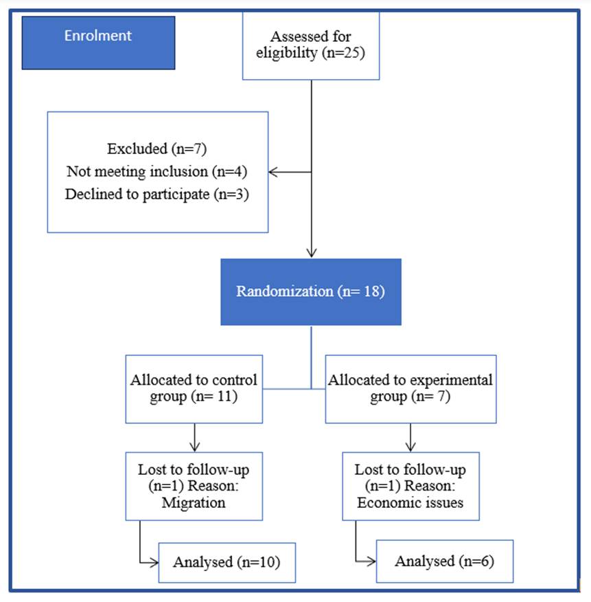
Flow chart 1: CONSORT diagram of the study
Informed consent was obtained from all subjects before participation. Participants were given detailed information about the purpose of the study, procedures involved, potential risks and benefits, and their right to withdraw at any stage. Information regarding the confidentiality of data was explained. Baseline demographic data were recorded.
Outcome measures included pain, range of motion, disability, muscle strength, and quality of life. Pain was assessed using the Visual Analogue Scale (VAS)[15]. It is a 10-point scale ranging from 0 (no pain) to 10 (worst imaginable pain). The range of motion of the shoulder joint was measured using a standard goniometer. The ranges of the following movements were assessed: flexion, extension, abduction, adduction, internal rotation and external rotation. Functional disability was assessed using the Disabilities of the Arm, Shoulder and Hand (DASH) questionnaire[16]. Higher DASH scores indicate a greater level of disability and symptom severity. Muscle strength was evaluated using the Modified Oxford Scale. The muscle groups assessed were shoulder flexors, extensors, abductors, adductors, internal rotators, and external rotators. The quality of life was assessed using the Short Form-36 (SF-36) questionnaire[17]. It includes eight domains: physical functioning, role limitations due to physical health, bodily pain, general health, vitality, social functioning, role limitations due to emotional health, and mental health. Scores range from 0 to 100, with higher scores indicating better health status.
Intervention was given to both groups for a duration of 6 weeks. Treatment sessions were conducted for 45 minutes, three times per week under supervision, and participants were instructed to continue exercises at home daily. Assessments were carried out at baseline (0 week), at the end of 3 weeks, and at the end of 6 weeks.
Group A (Control group) received conventional Occupational Therapy treatment based on standard clinical guidelines[13, 14]. This included cervical range of motion exercises, exercises and activities to increase shoulder joint range of motion, such as pendular exercises, shoulder pegs, shoulder pulley, inclined sanding, shoulder ladder and wand exercises. Also, ball squeezing to prevent complex regional pain syndrome. Adaptive training techniques included single-handed dressing technique, use of a dressing stick for upper extremity dressing, use of a long-handled body scrubber for bathing and reorganising frequently used items to waist-to-chest level countertop heights. Group B (Experimental group) received conventional Occupational Therapy along with additional scapulothoracic exercises. These included stretching exercises for levator scapulae, serratus anterior, rhomboids, and trapezius muscles; closed kinetic chain exercises such as wall push-ups and table push-ups; resistance band exercises including scapular extension and retraction; standing exercises involving scapular adduction, elevation, abduction, and stabilization using a therapy ball; supine scapular protraction; prone scapular adduction and extension; and shoulder isometric exercises.
Statistical Analysis:
Statistical Package for the Social Sciences (SPSS v 26.0, IBM) was used for the statistical analysis of the data. Descriptive statistics for numerical data included the mean with standard deviation (SD), and categorical data consisted of various parameters.
Descriptive statistics like frequencies and percentages for categorical data, Mean & SD for numerical data have
been depicted. Inter-group comparison for age (2 groups) was done using a t-test. Comparison of frequencies of categories of variables with groups was done using the chi-square test.
Normality of numerical data was checked using the Shapiro-Wilk test, and it was found that the data do not follow a normal curve; hence, non-parametric tests have been used for comparisons. Inter-group comparison (2 groups) was done using the Mann-Whitney U test. Intra-group comparison was done using the Wilcoxon Signed rank test (up to 2 observations) and using the Friedman test for more than 2 observations
RESULT
The mean age of participants was 53.69 ± 9.37 years. There was no statistically significant difference between Group A (53.40 ± 10.99 years) and Group B (54.17 ± 6.73 years) (p = 0.880), indicating homogeneity between groups.
Similarly, no significant differences were observed between the groups with respect to gender distribution (p = 0.091), side affected (p = 0.182), and hand dominance (p = 0.247), suggesting comparable baseline characteristics [Table. 1].
| Variable | Category | Group A (n=10) |
Group B (n=6) |
Total (n=16) |
Test |
p- |
|---|---|---|---|---|---|---|
| Age (years) | Mean ± SD | 53.40 ± 10.997 | 54.17 ± 6.735 | 53.69 ± 9.372 | t = -0.153 | 0.880 |
| Range | 31–67 | - | 31–67 | |||
| Gender | Female | 6 | 1 | 7 | χ² = 2.861 | 0.091 |
| Male | 4 | 5 | 9 | |||
| Diagnosis | Left Adhesive Capsulitis | 5 | 1 | 6 | χ² = 1.778 | 0.182 |
| Right Adhesive Capsulitis | 5 | 5 | 10 | |||
| Hand Dominance | Left | 1 | 2 | 3 | χ² = 1.340 | 0.247 |
| Right | 9 | 4 | 13 |
Table 1: Baseline characteristics of participants in the Control and Experimental groups
Group A is the control group, and Group B is the experimental group.
Both groups demonstrated significant improvement in all outcome measures over the 6-week intervention period. In the control group, significant improvements were observed in shoulder range of motion, pain intensity, disability scores, and quality of life domains from baseline to 6 weeks (p < 0.05) [Table. 2].
The experimental group demonstrated significant improvement in all assessed outcome measures over the 6-week intervention period. Significant gains were observed in shoulder range of motion, pain reduction, disability scores, muscle strength, and quality of life domains (p < 0.05) [Table. 3].
Both groups demonstrated improvement in range of motion, pain, and disability scores from baseline to 6 weeks. The experimental group showed comparatively greater improvement in shoulder ROM and DASH scores at 3 weeks and 6 weeks; however, the intergroup differences were not statistically significant (p > 0.05) [Table. 4].
| Outcome Measure |
Baseline (Mean±SD) |
3W (Mean±SD) |
6W (Mean±SD) |
Test | p- value |
|---|---|---|---|---|---|
| Flexion ROM | 110.50±24.20 | 120.00±25.49 | 127.50±26.38 | Friedman | <0.01 |
| Extension ROM | 41.00±15.42 | 45.50±13.63 | 51.00±9.66 | Friedman | <0.01 |
| Abduction ROM | 96.00±21.05 | 103.50±23.10 | 116.50±21.74 | Friedman | <0.01 |
| Internal Rotation | 51.00±14.29 | 55.50±12.57 | 59.00±12.87 | Friedman | <0.05 |
| External Rotation | 46.00±21.44 | 52.00±20.84 | 62.00±19.18 | Friedman | <0.01 |
| VAS | 4.30±1.49 | 3.40±1.84 | 3.10±1.52 | Friedman | <0.01 |
| DASH | 63.15±11.86 | 60.00±11.57 | 57.08±12.13 | Friedman | <0.01 |
| SF-36 Physical Function | 30.00±26.24 | 48.60±19.76 | 65.00±22.11 | Friedman | <0.01 |
| SF-36 Energy/Fatigue | 40.50±6.85 | 41.50±7.09 | 44.50±4.97 | Friedman | <0.01 |
| SF-36 Pain | 34.00±9.36 | 38.75±7.56 | 45.50±3.49 | Friedman | <0.01 |
| SF-36 General Health | 43.00±2.58 | 45.00±3.33 | 48.50±5.29 | Friedman | <0.01 |
Table 2: Intragroup comparison of Outcome Measures in control group A
ROM = Range of Motion
VAS = Visual Analogue Scale
DASH = Disabilities of the Arm, Shoulder and Hand
SF-36 = 36-item short-form survey
|
Outcome |
Baseline |
3W |
6W |
Test |
p- |
|---|---|---|---|---|---|
|
Flexion ROM |
98.33±19.14 |
121.67±21.37 |
140.00±19.75 |
Friedman |
<0.01 |
|
Extension ROM |
40.83±11.14 |
49.17±14.29 |
55.83±8.01 |
Friedman |
<0.01 |
|
Abduction ROM |
95.00±21.44 |
115.83±19.08 |
136.67±17.22 |
Friedman |
<0.01 |
|
Internal Rotation |
45.00±12.24 |
53.33±13.29 |
65.83±6.64 |
Friedman |
<0.01 |
|
External Rotation |
38.33±10.80 |
52.50±18.64 |
69.17±14.29 |
Friedman |
<0.01 |
|
VAS |
4.83±0.98 |
3.00±1.09 |
2.17±0.75 |
Friedman |
<0.01 |
|
DASH |
57.73±7.43 |
53.58±7.53 |
48.15±5.49 |
Friedman |
<0.01 |
|
SF-36 Physical Function |
41.67±21.37 |
68.33±11.25 |
91.67±12.91 |
Friedman |
<0.01 |
|
SF-36 Energy/Fatigue |
45.00±4.47 |
48.33±2.58 |
51.67±2.58 |
Friedman |
<0.05 |
|
SF-36 Pain |
32.92±7.14 |
44.58±5.57 |
53.75±8.33 |
Friedman |
<0.01 |
|
SF-36 General Health |
45.00±0.00 |
49.17±3.76 |
57.50±4.18 |
Friedman |
<0.01 |
Table 3: Intragroup comparison of Outcome Measures in experimental group B
ROM = Range of Motion
VAS = Visual Analogue Scale
DASH = Disabilities of the Arm, Shoulder and Hand
SF-36 = 36-item short-form survey
|
Outcome |
Timeline |
Group A |
Group B |
p- |
|---|---|---|---|---|
|
Flexion |
Baseline |
110.5±24.20 |
98.33±19.14 |
0.324 |
|
Flexion |
3 Weeks |
120.0±25.49 |
121.67±21.37 |
0.869 |
|
Flexion |
6 Weeks |
127.5±26.38 |
140.0±19.75 |
0.324 |
|
Extension |
Baseline |
41.0±15.42 |
40.83±11.14 |
0.869 |
|
Extension |
3 Weeks |
45.5±13.63 |
49.17±14.29 |
0.609 |
|
Extension |
6 Weeks |
51.0±9.66 |
55.83±8.01 |
0.433 |
|
Abduction |
Baseline |
96.0±21.05 |
95.0±21.44 |
0.870 |
|
Abduction |
3 Weeks |
103.5±23.10 |
115.83±19.08 |
0.352 |
|
Abduction |
6 Weeks |
116.5±21.74 |
136.67±17.22 |
0.071 |
|
Internal Rotation |
Baseline |
51.0±14.29 |
45.0±12.24 |
0.409 |
|
Internal Rotation |
3 Weeks |
55.5±12.57 |
53.33±13.29 |
0.740 |
|
Internal Rotation |
6 Weeks |
59.0±12.87 |
65.83±6.64 |
0.308 |
|
External Rotation |
Baseline |
46.0±21.44 |
38.33±10.80 |
0.701 |
|
External Rotation |
3 Weeks |
52.0±20.84 |
52.5±18.64 |
0.826 |
|
External Rotation |
6 Weeks |
62.0±19.18 |
69.17±14.29 |
0.376 |
|
VAS |
Baseline |
4.3±1.49 |
4.83±0.98 |
0.572 |
|
VAS |
3 Weeks |
3.4±1.84 |
3.0±1.09 |
0.619 |
|
VAS |
6 Weeks |
3.1±1.52 |
2.17±0.75 |
0.221 |
|
DASH |
Baseline |
63.15±11.87 |
57.73±7.44 |
0.301 |
|
DASH |
3 Weeks |
60.0±11.57 |
53.58±7.54 |
0.385 |
|
DASH |
6 Weeks |
57.08±12.14 |
48.15±5.49 |
0.115 |
Table 4: Intergroup comparison of Range of Motion, Pain and Disability outcomes
VAS = Visual Analogue Scale
DASH = Disabilities of the Arm, Shoulder and Hand
The experimental group B demonstrated significantly greater improvement in shoulder abductor muscle power and several SF-36 domains, including physical functioning, energy/fatigue, social functioning, pain, and general health at 3 weeks and 6 weeks (p < 0.05) as compared to control group A [Table. 5].
DISCUSSION
In this study, we evaluated the effect of a conventional Occupational Therapy program alone (Group A) against scapulothoracic exercises as an adjunct to the conventional Occupational Therapy program (Group B) in the treatment of adhesive capsulitis.
The findings demonstrated that both groups showed significant improvements in pain, range of motion (ROM), muscle strength, functional ability, and quality of life over
|
Parameter |
Timeline |
Group A |
Group B |
p- |
|---|---|---|---|---|
|
Abductors MP |
Baseline |
3.35±0.47 |
3.67±0.41 |
0.190 |
|
Abductors MP |
3 Weeks |
3.4±0.52 |
3.92±0.20 |
0.035* |
|
Abductors MP |
6 Weeks |
3.75±0.54 |
4.0±0.0 |
0.068 |
|
Physical Functioning |
Baseline |
30.0±26.25 |
41.67±21.37 |
0.420 |
|
Physical Functioning |
3 Weeks |
48.6±19.77 |
68.33±11.26 |
0.025* |
|
Physical Functioning |
6 Weeks |
65.0±22.11 |
91.67±12.91 |
0.024* |
|
Energy/Fatigue |
Baseline |
40.5±6.85 |
45.0±4.47 |
0.117 |
|
Energy/Fatigue |
3 Weeks |
41.5±7.09 |
48.33±2.58 |
0.023* |
|
Energy/Fatigue |
6 Weeks |
44.5±4.97 |
51.67±2.58 |
0.009* |
|
Social Function |
Baseline |
43.75±6.59 |
54.58±9.54 |
0.022* |
|
Social Function |
3 Weeks |
48.75±7.10 |
57.5±6.12 |
0.026* |
|
Social Function |
6 Weeks |
48.75±7.10 |
58.33±6.46 |
0.021* |
|
SF-36 Pain |
Baseline |
34.0±9.37 |
32.92±7.14 |
0.810 |
|
SF-36 Pain |
3 Weeks |
38.75±7.57 |
44.58±5.57 |
0.104 |
|
SF-36 Pain |
6 Weeks |
45.50±3.50 |
53.75±8.33 |
0.019* |
|
General Health |
Baseline |
43.0±2.58 |
45.0±0.0 |
0.083 |
|
General Health |
3 Weeks |
45.0±3.33 |
49.17±3.76 |
0.045* |
|
General Health |
6 Weeks |
48.5±5.30 |
57.5±4.18 |
0.008* |
Table 5: Intergroup comparison of Quality of Life and Muscle Power outcomes
SF-36 = 36-item short-form survey
*Significant difference seen if p values are less than 0.05, and highly significant difference seen if p values are less than 0.01
6 weeks. However, when compared, better results were seen in the experimental group in quality-of-life domains.
The significant intragroup improvements in both groups can be due to the effectiveness of the conventional rehabilitation program. The main focus of these interventions is on restoring glenohumeral joint mobility, reducing capsular tightness, and improving functional performance. Similar findings have been reported by Celik, whose study showed that structured exercise programs significantly improve ROM and functional outcomes in patients with frozen shoulder[1]. The improvements seen may be due to better capsular extensibility, enhanced synovial fluid dynamics, and a parallel decrease in pain-induced movement restriction.
The additional benefits observed in the experimental group may be attributed to the role of the scapula in shoulder biomechanics[2]. Changes in the scapular positions and movements, including increased protraction, abnormal rotation, and reduced posterior tilt, are commonly seen in adhesive capsulitis[3, 4]. These disturbances in the movement patterns may result in stress on surrounding structures.
In this study, scapulothoracic exercises likely contributed to improved neuromuscular control and coordination of scapular stabilisers such as the trapezius and serratus anterior. Brumitt emphasized the importance of early scapular stabilization exercises in restoring optimal shoulder function[5]. Similarly, Moezy et al. demonstrated that targeted scapular stabilization exercises significantly improve shoulder mobility, posture, and pain levels[6]. Scapular stabilization exercises may have resulted in better scapular positioning, leading to better co-ordination among the muscles, resulting in greater ROM as observed in the experimental group.
Pain reduction was observed in both groups, with relatively better outcomes in the experimental group. This may be due to the reduced abnormal mechanical stress and improved muscular balance around the shoulder complex. Cricchio and Frazer emphasised the importance of the scapulothoracic exercises in optimising muscle activation patterns and reducing compensatory overactivity, thereby decreasing pain[7]. Moreover, reduced muscle guarding may have contributed to the observed reduction in pain.
Although intergroup differences in ROM and muscle strength were not statistically significant at all time points, clinically meaningful improvements were evident in the experimental group. This may be due to the relatively small sample size and variability among participants. However, significant improvements in SF-36 domains such as physical functioning, energy/fatigue, social functioning, pain, and general health indicate a broader impact of scapulothoracic exercises on patient well-being.
The quality of life improvements observed in this study are consistent with previous research demonstrating a strong association between pain reduction, functional recovery, and enhanced quality of life in individuals with adhesive capsulitis[8, 9]. The inclusion of scapulothoracic exercises may have contributed to better participation in daily activities, thereby improving psychosocial aspects of health.
Previous evidence describes the significance of proximal stability in enhancing distal mobility. A study done by Kisner and Colby focuses on the role of coordinated muscle activity and kinetic chain function in efficient upper limb function[10]. Additionally, recent studies have shown that targeted exercise interventions of the scapulothoracic region can significantly improve ROM and functional outcomes in frozen shoulder[11]. A recent systematic review by Ferlito et al. also supports the inclusion of scapulothoracic interventions in the management of shoulder disorders, highlighting their effectiveness in improving function and reducing pain[12].
Despite these positive findings, certain limitations must be acknowledged. The sample size was small, which may limit the generalizability of the results. The duration of the study was limited to 6 weeks, and long-term outcomes were not assessed. Furthermore, a detailed biomechanical analysis of scapular movement was not performed, which could have provided additional insights into the mechanisms underlying the observed improvements.
CONCLUSION
In conclusion, the results of this study suggest that incorporating scapulothoracic exercises into conventional Occupational Therapy provides additional benefits in improving functional outcomes and quality of life in patients with adhesive capsulitis. These findings highlight the importance of addressing scapular mechanics as part of a comprehensive rehabilitation program.
DECLARATION
Acknowledgement: We would like to express our sincere gratitude to the Dean of Topiwala National Medical College and BYL Nair Hospital for allowing us to conduct this study.
Source of Funding: None.
Conflict of Interest: The authors declare no conflict of interest.
References
1. Celik D. Comparison of the outcomes of two different exercise programs on frozen shoulder. Acta Orthopaedica et Traumatologica Turcica. 2020; 44 (4). Available from: https://doi.org/10.3944/aott.2010.2367
2. Paine R, Voight ML. The role of the scapula. International Journal of Sports Physical Therapy. 2013; 8 (5). Available from: https://pubmed.ncbi.nlm.nih.gov/24175141/
3. Fayad F, Roby-Brami A, Yazbeck C, Hanneton S, Lefevre-Colau MM, Gautheron V, et al. Three-dimensional scapular kinematics and scapulohumeral rhythm in patients with glenohumeral osteoarthritis or frozen shoulder. Journal of Biomechanics. 2008; 41 (2). Available from: https://doi.org/10.1016/j.jbiomech.2007.09.004
4. Yano Y, Hamada J, Tamai K, Yoshizaki K, Sahara R, Fujiwara T, et al. Scapular kinematics during arm elevation. Journal of Shoulder and Elbow Surgery. 2010; 19 (2). Available from: https://doi.org/10.1016/j.jse.2009.09.007
5. Brumitt J. Scapular-stabilization exercises: early intervention prescription. Athletic Therapy Today. 2006; 11 (5). Available from: https://doi.org/10.1123/att.11.5.15
6. Moezy A, Sepehrifar S, Solaymani Dodaran M. The effects of scapular stabilization based exercise therapy on pain, posture, flexibility and shoulder mobility in patients with shoulder impingement syndrome: a controlled randomized clinical trial. Medical Journal of The Islamic Republic of Iran. 2014; 28 Available from: https://pubmed.ncbi.nlm.nih.gov/25664288/
7. Cricchio M, Frazer C. Scapulothoracic and Scapulohumeral Exercises: A Narrative Review of Electromyographic Studies. Journal of Hand Therapy. 2011; 24 (4). Available from: https://doi.org/10.1016/j.jht.2011.06.001
8. Bhagade RR, Sreeraj SR. Correlation between Pain, Functional Disability and Quality of Life with Sleep Disturbance in Patients with Adhesive Capsulitis. International Journal of Health Sciences and Research. 2018; 8 (6). Available from: https://www.ijhsr.org/IJHSR_Vol.8_Issue.6_June2018/19.pdf
9. Fernandes MR. Correlation between functional disability and quality of life in patients with adhesive capsulitis. Acta Ortopédica Brasileira. 2015; 23 (2). Available from: https://doi.org/10.1590/1413-78522015230200791
10. Kisner C, Colby LA. Therapeutic Exercise: Foundations and Techniques. 6th ed. Philadelphia: FA Davis; 2012.
11. Antari NK, Kinandana GP, Negara AA. Scapulothoracic joint mobilization improves outcomes in frozen shoulder. International Journal of Health Sciences and Research. 2021;9(3):166–175.
12. Ferlito R, Testa G, McCracken KL, Moscato S, Moscato GM, Panvini FMC, et al. Effectiveness of scapulothoracic interventions: systematic review. Journal of Functional Morphology and Kinesiology. 2023; 8 (2). Available from: https://doi.org/10.3390/jfmk8020038
13. Marik TL, Roll SC. Effectiveness of Occupational Therapy Interventions for Musculoskeletal Shoulder Conditions: A Systematic Review. American Journal of Occupational Therapy. 2017; 71 (1). Available from: https://doi.org/10.5014/ajot.2017.023127
14. Kelley MJ, Shaffer MA, Kuhn JE, Michener LA, Seitz AL, Uhl TL, et al. Shoulder Pain and Mobility Deficits: Adhesive Capsulitis. Journal of Orthopaedic & Sports Physical Therapy. 2013; 43 (5). Available from: https://doi.org/10.2519/jospt.2013.0302
15. Boonstra AM, Schiphorst Preuper HR, Reneman MF, Posthumus JB, Stewart RE. Reliability and validity of the visual analogue scale for disability in patients with chronic musculoskeletal pain. International Journal of Rehabilitation Research. 2008; 31 (2). Available from: https://doi.org/10.1097/mrr.0b013e3282fc0f93
16. Dixon D, Johnston M, McQueen M, Court-Brown C. The Disabilities of the Arm, Shoulder and Hand Questionnaire (DASH) can measure the impairment, activity limitations and participation restriction constructs from the International Classification of Functioning, Disability and Health (ICF). BMC Musculoskeletal Disorders. 2008; 9 (1). Available from: https://doi.org/10.1186/1471-2474-9-114
17. Lins L, Carvalho FM. SF-36 total score as a single measure of health-related quality of life: Scoping review. Sage Open Medicine. 2016; 4 Available from: https://doi.org/10.1177/2050312116671725
Copyright
©2026 Nagine & Koutarapu. This is an open-access journal, and articles are distributed under the terms of the Creative Commons Attribution License CC-BY 4.0. (https://creativecommons.org/licenses/by/4.0/) which permits unrestricted use, distribution, and reproduction in any medium, provided the original authors and source are credited.
Cite this article
Nagine SS, Koutarapu S. Effect of Scapulothoracic Exercises in the Treatment of Adhesive Capsulitis: A Randomized Controlled Trial. Perspectives in Medical Research. 2026; 14(2):85-91. DOI: 10.47799/pimr.1402.26.118