<?xml version='1.0' encoding='UTF-8'?>

<!DOCTYPE article PUBLIC "-//NLM//DTD JATS (Z39.96) Journal Publishing DTD v1.1d1 20130915//EN" "JATS-journalpublishing1.dtd">
<article xmlns:xlink="http://www.w3.org/1999/xlink">
  <front>
    <journal-meta id="journal-meta-87cddb9ab7774ac9973b6a64b7cbc767">
      <journal-id journal-id-type="nlm-ta">Sciresol</journal-id>
      <journal-id journal-id-type="publisher-id">Sciresol</journal-id>
      <journal-id journal-id-type="journal_submission_guidelines">https://jmsh.ac.in/</journal-id>
      <journal-title-group>
        <journal-title>Journal of Medical Sciences and Health</journal-title>
      </journal-title-group>
      <issn publication-format="print"/>
    </journal-meta>
    <article-meta>
        
          
            <article-id pub-id-type="doi">10.47799/pimr.1402.26.111</article-id>
          
          
            <article-categories>
              <subj-group>
                <subject>ORIGINAL ARTICLE</subject>
              </subj-group>
            </article-categories>
            <title-group>
              <article-title>&lt;p&gt;Histopathological Spectrum and Serum CA-125 Profile of Ovarian Neoplasms in a Malignancy-Predominant Surgical Cohort: A Retrospective Observational Study&lt;/p&gt;</article-title>
            </title-group>
          
          
            <pub-date date-type="pub">
              <day>30</day>
              <month>3</month>
              <year>2026</year>
            </pub-date>
            <permissions>
              <copyright-year>2026</copyright-year>
            </permissions>
          
          
            <volume>14</volume>
          
          
            <issue>2</issue>
          
          <fpage>1</fpage>

          <abstract>
            <title>Abstract</title>
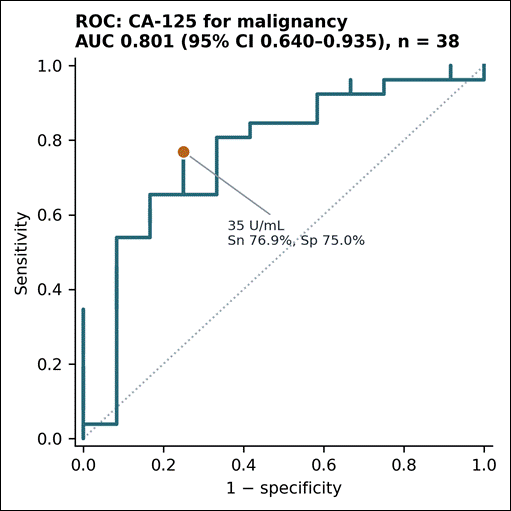
            &lt;p&gt;&lt;bold&gt;Background: &lt;/bold&gt;&lt;span&gt;Serum cancer antigen 125 (CA-125) is widely used in the evaluation and triage of adnexal masses. Several hospital-based Indian studies have reported very high diagnostic accuracy for CA-125 in distinguishing malignant from benign ovarian tumours. However, many such studies are derived from general gynaecological practice and contain predominantly benign lesions. The performance of CA-125 in malignancy-predominant cohorts, such as those encountered in tertiary oncology services, is less well characterised.&lt;/span&gt;&lt;bold&gt; Methods: &lt;/bold&gt;&lt;span&gt;We conducted a retrospective observational study of consecutively archived ovarian resection and oophorectomy specimens diagnosed at a single tertiary centre. Sixty unique patients were analysed after exclusion of duplicate records. Tumour behaviour and histogenetic category were assigned from the recorded histopathological diagnosis according to the fifth edition of the WHO Classification of Female Genital Tumours. Serum CA-125 values recorded at diagnosis were analysed using the conventional threshold of 35 U/mL. Because 45% of specimens were recorded as having been resected after neoadjuvant chemotherapy (NACT), a pre-specified sensitivity analysis was performed in patients without recorded NACT. Continuous variables were compared using the Mann–Whitney U test and categorical variables using Fisher exact test. Proportions are presented with exact 95% confidence intervals (CI), and diagnostic discrimination with the area under the receiver operating characteristic curve (AUC) and bootstrap 95% CI. No values were imputed. &lt;/span&gt;&lt;bold&gt;Results: &lt;/bold&gt;&lt;span&gt;Of 60 patients, 41 (68.3%had malignant tumours, 10 (16.7%) benign lesions, two (3.3%) borderline tumours, and seven (11.7%) nonspecific or unclassified lesions. Epithelial tumours predominated (46/60, 76.7%). Median age was 46.0 years (IQR 42.0–56.0) and did not differ between malignant and benign groups (p=0.716). CA-125 was available for 38 patients and was significantly higher in malignant than benign tumours (median 100.6 vs 23.2 U/mL; p=0.0069). At 35 U/mL, sensitivity was 76.9% (95% CI 56.4–91.0), specificity 75.0% (42.8–94.5), and AUC 0.801 (0.640–0.935). Six of 26 malignancies had CA-125 values below 35 U/mL. In patients without recorded NACT, sensitivity decreased to 63.6% and AUC to 0.727. Among malignant tumours, CA-125 was higher in specimens following NACT than in those without recorded NACT (median 420.3 vs 46.2 U/mL; p=0.049). Bilateral ovarian involvement was significantly associated with malignancy (12/17 malignant vs 1/9 benign/borderline; p=0.011). &lt;/span&gt;&lt;bold&gt;Conclusions: &lt;/bold&gt;&lt;span&gt;In this malignancy-predominant cohort, CA-125 at the conventional 35 U/mL threshold showed only moderate discrimination and missed approximately one-quarter of malignancies. Bilaterality demonstrated a stronger association with malignancy than the CA-125 threshold. The findings suggest that the near-perfect performance reported in some benign-predominant surgical series may partly reflect case mix. CA-125 should therefore not be interpreted in isolation in referral populations. Prospective studies incorporating marker timing, menopausal status, and imaging morphology are required to evaluate its performance in multimodal diagnostic models.&lt;/span&gt;&lt;/p&gt;
          </abstract>
          
          
            <kwd-group>
              <title>Keywords</title>
              
                <kwd>Ovarian neoplasms; CA-125; Histopathology; Diagnostic accuracy; Neoadjuvant chemotherapy; India</kwd>
              
            </kwd-group>


        <contrib-group>
          
            
              <contrib contrib-type="author">
                <name>
                  <surname>Dharwar</surname>
                  <given-names>Amarendra</given-names>
                </name>
                
                  <xref rid="aff-1" ref-type="aff">1</xref>
                
              </contrib>


              <aff id="aff-1">
                <institution> Department of Pathology / Obstetrics and Gynaecology VTSM Peripheral Cancer Centre, Kidwai Institute of Oncology </institution>
                <addr-line>Kalaburagi, Karnataka India</addr-line>
              </aff>
            
              <aff id="aff-2">
                <institution> Assistant professor, Pathology VTSM Peripheral Cancer Centre, Kidwai Institute of Oncology </institution>
                <addr-line>Kalaburagi, Karnataka India</addr-line>
              </aff>


              <contrib contrib-type="author">
                <name>
                  <surname>Sana</surname>
                  <given-names>Sumaiyya</given-names>
                </name>
                
                  <xref rid="aff-1" ref-type="aff">1</xref>
                
              </contrib>


              <aff id="aff-1">
                <institution> Department of Pathology / Obstetrics and Gynaecology VTSM Peripheral Cancer Centre, Kidwai Institute of Oncology </institution>
                <addr-line>Kalaburagi, Karnataka India</addr-line>
              </aff>
            
              <aff id="aff-2">
                <institution> Assistant professor, Pathology VTSM Peripheral Cancer Centre, Kidwai Institute of Oncology </institution>
                <addr-line>Kalaburagi, Karnataka India</addr-line>
              </aff>


              <contrib contrib-type="author">
                <name>
                  <surname>Farheen</surname>
                  <given-names>Javeriya</given-names>
                </name>
                
                  <xref rid="aff-2" ref-type="aff">2</xref>
                
              </contrib>


              <aff id="aff-1">
                <institution> Department of Pathology / Obstetrics and Gynaecology VTSM Peripheral Cancer Centre, Kidwai Institute of Oncology </institution>
                <addr-line>Kalaburagi, Karnataka India</addr-line>
              </aff>
            
              <aff id="aff-2">
                <institution> Assistant professor, Pathology VTSM Peripheral Cancer Centre, Kidwai Institute of Oncology </institution>
                <addr-line>Kalaburagi, Karnataka India</addr-line>
              </aff>


              <contrib contrib-type="author">
                <name>
                  <surname>Aziz</surname>
                  <given-names>Abu Hasim Abdul</given-names>
                </name>
                
                  <xref rid="aff-1" ref-type="aff">1</xref>
                
              </contrib>


              <aff id="aff-1">
                <institution> Department of Pathology / Obstetrics and Gynaecology VTSM Peripheral Cancer Centre, Kidwai Institute of Oncology </institution>
                <addr-line>Kalaburagi, Karnataka India</addr-line>
              </aff>
            
              <aff id="aff-2">
                <institution> Assistant professor, Pathology VTSM Peripheral Cancer Centre, Kidwai Institute of Oncology </institution>
                <addr-line>Kalaburagi, Karnataka India</addr-line>
              </aff>
            
          
        </contrib-group>
        
    </article-meta>
  </front>
  <body>
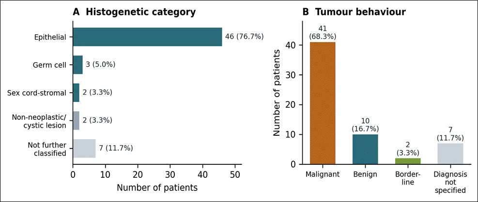
    <heading><span><bold>INTRODUCTION</bold></span></heading><p><span>Ovarian cancer remains an important cause of cancer morbidity and mortality worldwide. In India, a substantial proportion of patients present with advanced disease, reflecting the often asymptomatic nature of early ovarian cancer and the deep pelvic location of the ovaries<superscript>[<xref ref-type="link" rid="#ref-1">1</xref>-<xref ref-type="link" rid="#ref-3">3</xref>]</superscript>. Preoperative discrimination between benign and malignant adnexal masses is consequently important for appropriate referral, surgical planning, and selection between primary cytoreductive surgery and neoadjuvant chemotherapy (NACT) followed by interval debulking surgery.</span></p><p><span>Serum cancer antigen 125 (CA-125), a MUC16-derived glycoprotein, remains the most widely available serum biomarker used in the evaluation of suspected ovarian malignancy, with 35 U/mL being the conventional clinical threshold<superscript>[<xref ref-type="link" rid="#ref-4">4</xref>]</superscript>. However, CA-125 is not specific for ovarian malignancy and may be elevated in benign gynaecological and non-gynaecological conditions. Conversely, normal concentrations occur in early-stage disease and in several non-serous histological subtypes <superscript>[<xref ref-type="link" rid="#ref-5">5</xref>, <xref ref-type="link" rid="#ref-6">6</xref>]</superscript>. Population-based evidence has therefore demonstrated more modest diagnostic performance than might be expected from selected surgical series<superscript>[<xref ref-type="link" rid="#ref-7">7</xref>, <xref ref-type="link" rid="#ref-8">8</xref>]</superscript>.</span></p><p><span>Several recent Indian hospital-based studies have nevertheless reported sensitivities approaching 100% and areas under the receiver operating characteristic curve (AUC) exceeding 0.95. These studies frequently contain large numbers of unequivocally benign ovarian lesions compared with relatively few advanced cancers. Such case composition may exaggerate apparent diagnostic discrimination because the malignant and benign groups represent biological extremes.</span></p><p><span>The present study therefore evaluated the histopathological spectrum of ovarian lesions in a consecutive archival surgical cohort from a tertiary centre and assessed the discriminatory performance of CA-125 at the conventional 35 U/mL threshold. Particular attention was given to the potential influence of NACT on the interpretation of recorded CA-125 values.</span></p><heading><span><bold>MATERIALS AND METHODS</bold></span></heading><p><span><bold>Study design and participants</bold></span></p><p><span>This was a single-centre retrospective observational study conducted in the Department of Pathology, VTSM PCC, Kalaburagi. Cases were identified from the departmental histopathology archive. All patients with ovarian resection or oophorectomy specimens receiving a histopathological diagnosis during the available study period were eligible. No exclusion was made according to age, tumour behaviour, or availability of CA-125.</span></p><p><span>Sixty-three register entries were initially identified. Three duplicate records relating to the same patients were excluded, leaving 60 unique patients. The cohort represented consecutive available cases, with no a priori sample-size calculation.</span></p><p><span><bold>Histopathological classification and variables</bold></span></p><p><span>The histopathological diagnosis recorded in the pathology report was used as the reference standard. Tumour behaviour was categorised as benign, borderline, or malignant, and histogenetic category was assigned according to the fifth edition of the WHO Classification of Female Genital Tumours<superscript>[<xref ref-type="link" rid="#ref-9">9</xref>, <xref ref-type="link" rid="#ref-10">10</xref>]</superscript>. Diagnoses containing “carcinoma” or “yolk sac tumour” were classified as malignant, whereas diagnoses containing “borderline” were classified as borderline. Records containing only a nonspecific description such as “ovarian tumour” were retained descriptively but excluded from behaviour-specific comparisons.</span></p><p><span>Laterality, pathological T category, FIGO stage, NACT status, and omental/peritoneal findings were abstracted where recorded. NACT status was considered positive only when explicitly documented; an unrecorded treatment field was not assumed to indicate primary surgery.</span></p><p><span><bold>CA-125 analysis</bold></span></p><p><span>Serum CA-125 was analysed both as a continuous variable and dichotomised at 35 U/mL. The recorded value was used without imputation. Borderline tumours were grouped with non-malignant lesions for calculation of diagnostic performance.</span></p><p><span>Because the source register did not consistently document the timing of CA-125 sampling relative to chemotherapy, a sensitivity analysis was performed among patients without NACT recorded. The assay platform and reference interval could not be recovered from the archived records.</span></p><p><span><bold>Statistical analysis</bold></span></p><p><span>Continuous variables were summarised using mean ± standard deviation and median with interquartile range (IQR). CA-125 was reported primarily using median and IQR because of its right-skewed distribution. Two-group continuous comparisons used the Mann–Whitney U test with the Hodges–Lehmann estimator and 95% CI. Categorical variables were compared using Fisher exact test. Sensitivity, specificity, positive predictive value (PPV), negative predictive value (NPV), and accuracy were calculated for the 35 U/mL threshold. Exact Clopper–Pearson 95% CIs were used for proportions. The AUC was estimated using 10,000 bootstrap resamples with a percentile 95% CI.</span></p><p><span>No multivariable model was fitted because the cohort contained only 10 benign comparators and would not support reliable modelling. Two-sided p&lt;0.05 was considered statistically significant, although all analyses were regarded as exploratory because of the sample size and multiple comparisons.</span></p><heading><span><bold>RESULTS</bold></span></heading><p><span><bold>Cohort characteristics and histopathological spectrum</bold></span></p><p><span>Of 60 unique patients, 59 (98.3%) had a recorded histopathological diagnosis. A specific histological subtype was documented in 37 (61.7%), CA-125 in 38 (63.3%), laterality in 26 (43.3%), FIGO stage in 17 (28.3%), and NACT status in 27 (45.0%).</span></p><p><span>Malignant tumours accounted for 41/60 cases (68.3%; 95% CI 55.0–79.7), while 10 (16.7%; 8.3–28.5) were benign and two (3.3%; 0.4–11.5) borderline. Seven cases (11.7%; 4.8–22.6) had insufficiently specific documentation to assign behaviour. Epithelial tumours predominated (46/60, 76.7%), followed by germ cell tumours (3/60, 5.0%) and sex cord–stromal tumours (2/60, 3.3%).</span></p><figure id="table-1"><table><thead><tr><th><span><bold>Variable</bold></span></th><th><span><bold>Recorded, n</bold></span></th><th><span><bold>% of cohort</bold></span></th></tr></thead><tbody><tr><td><span>Age</span></td><td><span>57</span></td><td><span>95.0</span></td></tr><tr><td><span>Histopathological diagnosis (any)</span></td><td><span>59</span></td><td><span>98.3</span></td></tr><tr><td><span>Histological subtype specified</span></td><td><span>37</span></td><td><span>61.7</span></td></tr><tr><td><span>Serum CA-125 concentration</span></td><td><span>38</span></td><td><span>63.3</span></td></tr><tr><td><span>Laterality</span></td><td><span>26</span></td><td><span>43.3</span></td></tr><tr><td><span>Pathological (pT) category</span></td><td><span>18</span></td><td><span>30.0</span></td></tr><tr><td><span>FIGO stage</span></td><td><span>17</span></td><td><span>28.3</span></td></tr><tr><td><span>Neoadjuvant chemotherapy recorded</span></td><td><span>27</span></td><td><span>45.0</span></td></tr><tr><td><span>Omental / peritoneal findings</span></td><td><span>18</span></td><td><span>30.0</span></td></tr></tbody></table><figcaption><span><bold>Table 1: Completeness of recorded variables (N = 60)</bold></span></figcaption></figure><p> </p><figure id="table-2"><table><thead><tr><th><span><bold>Category</bold></span></th><th><span><bold>n</bold></span></th><th><span><bold>%</bold></span></th><th><span><bold>95% CI (%)</bold></span></th></tr></thead><tbody><tr><td><span>Behaviour</span></td><td> </td><td> </td><td> </td></tr><tr><td><span>  Malignant</span></td><td><span>41</span></td><td><span>68.3</span></td><td><span>55.0–79.7</span></td></tr><tr><td><span>  Benign</span></td><td><span>10</span></td><td><span>16.7</span></td><td><span>8.3–28.5</span></td></tr><tr><td><span>  Borderline</span></td><td><span>2</span></td><td><span>3.3</span></td><td><span>0.4–11.5</span></td></tr><tr><td><span>  Not further specified</span></td><td><span>7</span></td><td><span>11.7</span></td><td><span>4.8–22.6</span></td></tr><tr><td><span>Histogenetic category</span></td><td> </td><td> </td><td> </td></tr><tr><td><span>  Epithelial</span></td><td><span>46</span></td><td><span>76.7</span></td><td><span>—</span></td></tr><tr><td><span>  Germ cell</span></td><td><span>3</span></td><td><span>5.0</span></td><td><span>—</span></td></tr><tr><td><span>  Sex cord–stromal</span></td><td><span>2</span></td><td><span>3.3</span></td><td><span>—</span></td></tr><tr><td><span>  Non-neoplastic / cystic lesion</span></td><td><span>2</span></td><td><span>3.3</span></td><td><span>—</span></td></tr><tr><td><span>  Not further classified</span></td><td><span>7</span></td><td><span>11.7</span></td><td><span>—</span></td></tr></tbody></table><figcaption><span><bold>Table 2: Tumour behaviour and histogenetic category (N = 60)</bold></span></figcaption></figure><p><italic><span>CI, confidence interval (Clopper–Pearson exact). Confidence intervals are given for the behaviour categories, which constitute the primary descriptive outcome.</span></italic></p><p> </p><p><span>Among specifically recorded diagnoses, high-grade serous carcinoma and high-grade serous papillary carcinoma were the most frequent malignant categories, with nine cases each (15.0%). Mucinous carcinoma accounted for four cases (6.7%). Thirteen malignancies (21.7%) were recorded only as “carcinoma ovary” without further subtype. Mucinous cystadenoma was the most frequent benign neoplasm (3/60, 5.0%).</span></p><figure id="figure-1"><graphic src="https://schoproductionportal.s3.ap-south-1.amazonaws.com/data/PIMR/445/1787894146393.png"/><figcaption><span><bold>Fig. 1: Distribution of ovarian lesions by histogenetic category (A) and tumour behaviour (B), N = 60</bold></span></figcaption></figure><p> </p><figure id="table-3"><table><thead><tr><th><p><span><bold>Reported diagnosis</bold></span></p></th><th><p><span><bold>n</bold></span></p></th><th><p><span><bold>%</bold></span></p></th></tr></thead><tbody><tr><td><p><span>Carcinoma, subtype not specified</span></p></td><td><p><span>13</span></p></td><td><p><span>21.7</span></p></td></tr><tr><td><p><span>High-grade serous carcinoma</span></p></td><td><p><span>9</span></p></td><td><p><span>15.0</span></p></td></tr><tr><td><p><span>High-grade serous papillary carcinoma</span></p></td><td><p><span>9</span></p></td><td><p><span>15.0</span></p></td></tr><tr><td><p><span>Recorded only as "ovarian tumour"</span></p></td><td><p><span>6</span></p></td><td><p><span>10.0</span></p></td></tr><tr><td><p><span>Mucinous carcinoma</span></p></td><td><p><span>4</span></p></td><td><p><span>6.7</span></p></td></tr><tr><td><p><span>Mucinous cystadenoma</span></p></td><td><p><span>3</span></p></td><td><p><span>5.0</span></p></td></tr><tr><td><p><span>Mature cystic teratoma</span></p></td><td><p><span>2</span></p></td><td><p><span>3.3</span></p></td></tr><tr><td><p><span>Serous carcinoma, grade unspecified</span></p></td><td><p><span>2</span></p></td><td><p><span>3.3</span></p></td></tr><tr><td><p><span>Poorly differentiated carcinoma</span></p></td><td><p><span>2</span></p></td><td><p><span>3.3</span></p></td></tr><tr><td><p><span>Mucinous cystadenocarcinoma</span></p></td><td><p><span>1</span></p></td><td><p><span>1.7</span></p></td></tr><tr><td><p><span>Borderline mucinous tumour</span></p></td><td><p><span>1</span></p></td><td><p><span>1.7</span></p></td></tr><tr><td><p><span>Borderline cystadenoma, type not specified</span></p></td><td><p><span>1</span></p></td><td><p><span>1.7</span></p></td></tr><tr><td><p><span>Yolk sac tumour</span></p></td><td><p><span>1</span></p></td><td><p><span>1.7</span></p></td></tr><tr><td><p><span>Fibroma</span></p></td><td><p><span>1</span></p></td><td><p><span>1.7</span></p></td></tr><tr><td><p><span>Cellular fibroma</span></p></td><td><p><span>1</span></p></td><td><p><span>1.7</span></p></td></tr><tr><td><p><span>Serous cystadenoma</span></p></td><td><p><span>1</span></p></td><td><p><span>1.7</span></p></td></tr><tr><td><p><span>Benign ovarian cyst</span></p></td><td><p><span>1</span></p></td><td><p><span>1.7</span></p></td></tr><tr><td><p><span>Haemorrhagic cyst</span></p></td><td><p><span>1</span></p></td><td><p><span>1.7</span></p></td></tr><tr><td><p><span>Diagnosis not recorded</span></p></td><td><p><span>1</span></p></td><td><p><span>1.7</span></p></td></tr></tbody></table><figcaption><span><bold>Table 3: Histological subtypes as recorded (N = 60)</bold></span></figcaption></figure><heading> </heading><p><span><bold>Age and laterality</bold></span></p><p><span>Age was available for 57 patients (95.0%). The median age was 46.0 years (IQR 42.0–56.0; range 12–75). The largest age group was 41–50 years (23/60, 38.3%). Median age did not differ significantly between malignant and benign groups: 46.5 versus 44.0 years, respectively (Hodges–Lehmann difference 2.0 years, 95% CI −9.0 to 13.0; p=0.716). Laterality was recorded in 26 patients. Bilateral involvement occurred in 12/17 malignant tumours compared with 1/9 benign or borderline lesions (Fisher exact p=0.011). Right-sided unilateral disease was recorded in five of nine benign/borderline lesions compared with two of 17 malignant tumours.</span></p><figure id="table-4"><table><thead><tr><th><p><span><bold>Age group (years)</bold></span></p></th><th><p><span><bold>n</bold></span></p></th><th><p><span><bold>% of cohort</bold></span></p></th><th> </th></tr></thead><tbody><tr><td><p><span>≤20</span></p></td><td><p><span>2</span></p></td><td><p><span>3.3</span></p></td><td> </td></tr><tr><td><p><span>21–30</span></p></td><td><p><span>2</span></p></td><td><p><span>3.3</span></p></td><td> </td></tr><tr><td><p><span>31–40</span></p></td><td><p><span>9</span></p></td><td><p><span>15.0</span></p></td><td> </td></tr><tr><td><p><span>41–50</span></p></td><td><p><span>23</span></p></td><td><p><span>38.3</span></p></td><td> </td></tr><tr><td><p><span>51–60</span></p></td><td><p><span>14</span></p></td><td><p><span>23.3</span></p></td><td> </td></tr><tr><td><p><span>61–70</span></p></td><td><p><span>5</span></p></td><td><p><span>8.3</span></p></td><td> </td></tr><tr><td><p><span>71–80</span></p></td><td><p><span>2</span></p></td><td><p><span>3.3</span></p></td><td> </td></tr><tr><td><p><span>Not recorded</span></p></td><td><p><span>3</span></p></td><td><p><span>5.0</span></p></td><td> </td></tr><tr><td><p><span><bold>Behaviour</bold></span></p></td><td><p><span><bold>n with </bold></span><line-break/><span><bold>age</bold></span></p></td><td><p><span><bold>Mean ± SD </bold></span><line-break/><span><bold>(years)</bold></span></p></td><td><p><span><bold>Median </bold></span><line-break/><span><bold>(IQR)</bold></span></p></td></tr><tr><td><p><span>Benign</span></p></td><td><p><span>10</span></p></td><td><p><span>46.8 ± 17.6</span></p></td><td><p><span>44.0 (36.2–59.2)</span></p></td></tr><tr><td><p><span>Borderline</span></p></td><td><p><span>1</span></p></td><td><p><span>—</span></p></td><td><p><span>62.0</span></p></td></tr><tr><td><p><span>Malignant</span></p></td><td><p><span>40</span></p></td><td><p><span>48.0 ± 11.0</span></p></td><td><p><span>46.5 (42.0–55.2)</span></p></td></tr><tr><td><p><span>Not further specified</span></p></td><td><p><span>6</span></p></td><td><p><span>43.3 ± 14.0</span></p></td><td><p><span>45.0 (38.5–51.5)</span></p></td></tr></tbody></table><figcaption><span><bold>Table 4: Age distribution and age by tumour behaviour</bold></span></figcaption></figure><p><italic><span>SD, standard deviation; IQR, interquartile range. Malignant versus benign: Mann–Whitney U = 215.5, p = 0.716.</span></italic></p><p> </p><figure id="table-5"><table><thead><tr><th><span><bold>Laterality</bold></span></th><th><span><bold>Benign / borderline (n = 9)</bold></span></th><th><span><bold>Malignant (n = 17)</bold></span></th><th><span><bold>Total</bold></span></th></tr></thead><tbody><tr><td><span>Bilateral</span></td><td><span>1</span></td><td><span>12</span></td><td><span>13</span></td></tr><tr><td><span>Right ovary only</span></td><td><span>5</span></td><td><span>2</span></td><td><span>7</span></td></tr><tr><td><span>Left ovary only</span></td><td><span>3</span></td><td><span>3</span></td><td><span>6</span></td></tr></tbody></table><figcaption><span><bold>Table 5: Laterality by tumour behaviour, among the 26 patients with laterality recorded</bold></span></figcaption></figure><p><italic><span>Bilateral versus unilateral, malignant versus benign/borderline: Fisher exact p = 0.011. The odds ratio (19.2) is presented for completeness only; with a single bilateral benign case its confidence interval is uninformative, and it should not be interpreted as an effect estimate.</span></italic></p><p> </p><p><span><bold>Laterality</bold></span></p><p><span>Laterality was documented for 26 patients (43.3%). Among these, bilateral involvement was present in 13 (50.0%), the right ovary alone in seven (26.9%), and the left ovary alone in six (23.1%). Bilaterality was strongly associated with malignancy: 12 of 17 malignant tumours with recorded laterality were bilateral, compared with one of nine benign or borderline lesions (Fisher exact p = 0.011; <xref ref-type="link" rid="#table-5">[Table. 5]</xref>). Conversely, unilateral right-sided disease was more characteristic of benign lesions (five of nine) than of malignancy (two of 17).</span></p><p><span><bold>Serum CA-125</bold></span></p><p><span>CA-125 was available in 38 patients. The overall median concentration was 45.9 U/mL (IQR 25.4–212.1; range 6.0–3457.0). Malignant tumours had significantly higher CA-125 concentrations than benign lesions (median 100.6 vs 23.2 U/mL; Hodges–Lehmann difference 55.1 U/mL, 95% CI 10.0–390.7; p=0.0069).</span></p><p><span>At the 35 U/mL threshold, 20/26 malignant tumours (76.9%; 95% CI 56.4–91.0) and 3/10 benign lesions (30.0%; 6.7–65.2) had elevated CA-125 (Fisher exact p=0.004). Six malignant tumours had CA-125 below 35 U/mL. These included one high-grade serous carcinoma (7.3 U/mL), one mucinous carcinoma (20.4 U/mL), one yolk sac tumour (26.9 U/mL), one high-grade serous papillary carcinoma (29.5 U/mL), and two carcinomas without further subtype (12.6 and 21.7 U/mL). The three benign lesions with elevated CA-125 were two mucinous cystadenomas and one serous cystadenoma.</span></p><p> </p><figure id="table-6"><table><thead><tr><th><span><bold>Behaviour</bold></span></th><th><span><bold>n</bold></span></th><th><span><bold>Median (IQR), U/mL</bold></span></th><th><span><bold>Range, U/mL</bold></span></th><th><span><bold>&gt;35 U/mL, n (%)</bold></span></th><th><span><bold>95% CI (%)</bold></span></th></tr></thead><tbody><tr><td><span>Benign</span></td><td><span>10</span></td><td><span>23.2 (10.8–41.6)</span></td><td><span>6.0–214.0</span></td><td><span>3 (30.0)</span></td><td><span>6.7–65.2</span></td></tr><tr><td><span>Borderline</span></td><td><span>2</span></td><td><span>27.0 (26.0–27.9)</span></td><td><span>25.1–28.8</span></td><td><span>0 (0.0)</span></td><td><span>0.0–84.2</span></td></tr><tr><td><span>Malignant</span></td><td><span>26</span></td><td><span>100.6 (36.2–444.3)</span></td><td><span>7.3–3457.0</span></td><td><span>20 (76.9)</span></td><td><span>56.4–91.0</span></td></tr><tr><td><span>All</span></td><td><span>38</span></td><td><span>45.9 (25.4–212.1)</span></td><td><span>6.0–3457.0</span></td><td><span>23 (60.5)</span></td><td><span>43.4–76.0</span></td></tr></tbody></table><figcaption><span><bold>Table 6: Serum CA-125 by tumour behaviour, among the 38 patients with a recorded value</bold></span></figcaption></figure><p><italic><span>IQR, interquartile range; CI, confidence interval (Clopper–Pearson exact). Malignant versus benign: Mann–Whitney U = 207.0, p = 0.0069. Proportion above threshold across the three behaviour groups: Fisher exact p = 0.004.</span></italic></p><p> </p><p><span><bold>Diagnostic performance</bold></span></p><p><span>When borderline tumours were classified as non-malignant, CA-125 &gt;35 U/mL yielded 20 true-positive, six false-negative, three false-positive, and nine true-negative results.</span></p><figure><table><tbody><tr><td><span>Positive predictive </span><line-break/><span>value</span></td><td><span>87.0%</span></td><td><span>66.4–97.2</span></td><td><span>70.0%</span></td><td><span>34.8–93.3</span></td></tr><tr><td><span>Negative predictive </span><line-break/><span>value</span></td><td><span>60.0%</span></td><td><span>32.3–83.7</span></td><td><span>69.2%</span></td><td><span>38.6–90.9</span></td></tr><tr><td><span>Accuracy</span></td><td><span>76.3%</span></td><td><span>59.8–88.6</span></td><td><span>69.6%</span></td><td><span>47.1–86.8</span></td></tr><tr><td><span>Area under </span><line-break/><span>ROC curve</span></td><td><span>0.801</span></td><td><span>0.640–0.935</span></td><td><span>0.727</span></td><td><span>not estimated</span></td></tr></tbody></table></figure><p> </p><p><span>Sensitivity was 76.9% (95% CI 56.4–91.0), specificity 75.0% (42.8–94.5), PPV 87.0% (66.4–97.2), NPV 60.0% (32.3–83.7), and accuracy 76.3% (59.8–88.6). The AUC for CA-125 as a continuous variable was 0.801 (bootstrap 95% CI 0.640–0.935).</span></p><p><span>The predictive values reflect the high prevalence of malignancy in this cohort and therefore should not be extrapolated to lower-prevalence general gynaecological populations.</span></p><figure id="figure-2"><graphic src="https://schoproductionportal.s3.ap-south-1.amazonaws.com/data/PIMR/445/1787894146414.png"/><figcaption><span><bold>Fig. 2: Receiver operating characteristic curve for serum CA-125 in the discrimination of malignant from benign and borderline ovarian tumours (n = 38). The marked point corresponds to the conventional 35 U/mL threshold</bold></span></figcaption></figure><p> </p><p><span><bold>NACT, stage and peritoneal findings</bold></span></p><p><span>Twenty-seven specimens (45.0%) were recorded as following NACT. Among malignant tumours, CA-125 was significantly higher in patients whose specimens followed NACT than in those without NACT recorded (median 420.3 vs 46.2 U/mL; p=0.049).</span></p><p><span>In the sensitivity analysis restricted to 23 patients without recorded NACT, sensitivity decreased from 76.9% to 63.6% (95% CI 30.8–89.1), while specificity remained 75.0% and the AUC decreased to 0.727.</span></p><p><span>FIGO stage was available for only 17 patients: seven stage I, one stage II, eight stage III, and one stage IV. Median CA-125 was higher in stage III–IV than stage I–II disease (206.4 vs 69.7 U/mL), although the difference was not statistically significant (p=0.074). Omental or ascitic findings were recorded in 18 patients. No significant association between CA-125 and omental deposits was identified (p=0.797).</span></p><heading><span><bold>DISCUSSION</bold></span></heading><p><span>This retrospective series describes 60 ovarian resection specimens in which malignancy predominated, accounting for more than two-thirds of cases. The central observation is that within such a cohort, serum CA-125 at the conventional 35 U/mL threshold discriminated malignant from benign disease only moderately — sensitivity 76.9%, specificity 75.0%, AUC 0.801 — and missed approximately one malignancy in four. Restricting the analysis to patients with no recorded neoadjuvant chemotherapy, which is the population in which a preoperative marker is actually deployed, reduced sensitivity further to 63.6%.</span></p><p><span>This performance sits well below that reported by several recent Indian histopathological series, some of which describe sensitivities of 100% and areas under the curve exceeding 0.99. The discrepancy is unlikely to reflect assay differences and is more plausibly explained by case mix and by the composition of the comparator group. In benign-predominant series, the malignant group is typically dominated by advanced high-grade serous carcinoma with markedly elevated markers, while the non-malignant group consists largely of unequivocally benign, marker-negative lesions; discrimination between these two extremes is close to trivial. Our cohort contained six marker-negative malignancies, including a yolk sac tumour and a mucinous carcinoma — histotypes in which CA-125 is well recognised to be insensitive<superscript>[<xref ref-type="link" rid="#ref-4">4</xref>, <xref ref-type="link" rid="#ref-6">6</xref>]</superscript> — and three marker-positive benign lesions, two of them mucinous cystadenomas. Both patterns are entirely consistent with the established biology of the marker and with primary-care and registry-based evidence that CA-125 has moderate rather than near-perfect discrimination<superscript>[<xref ref-type="link" rid="#ref-7">7</xref>, <xref ref-type="link" rid="#ref-8">8</xref>]</superscript>. Reported accuracy figures from single-centre surgical series should therefore be read as properties of the case mix as much as of the test.</span></p><p><span>The predictive values we report illustrate the same point from the opposite direction. With 68% malignancy prevalence, a positive CA-125 carried a positive predictive value of 87%, while a normal result reduced the probability of malignancy only to 40%. In a general gynaecological population, where malignancy prevalence is closer to 20%, the same sensitivity and specificity would generate a far lower positive predictive value and a far higher negative predictive value. Predictive values are not properties of the assay, and their uncritical transfer between settings is a recurring weakness in this literature.</span></p><p><span>The association between bilateral ovarian involvement and malignancy was the strongest signal in our data: 12 of 17 malignancies with documented laterality were bilateral, against one of nine benign or borderline lesions. This accords with the recognised propensity of high-grade serous carcinoma, which comprised the largest specific subtype here, to involve both ovaries, and it is a reminder that morphological features available at imaging may carry more discriminatory weight in a referral setting than a single marker threshold. The finding must be treated cautiously: laterality was documented for fewer than half the cohort, and its recording may itself have been more thorough in oncological cases.</span></p><p><span>Our finding that CA-125 was substantially higher among malignancies whose specimen followed neoadjuvant chemotherapy is best read as a marker of disease extent rather than of treatment effect, since patients triaged to NACT are by definition those with bulky or unresectable disease. Baseline CA-125 has repeatedly been shown to relate to tumour burden and to the likelihood of complete cytoreduction, and CA-125 kinetics during chemotherapy carry independent prognostic information<superscript>[<xref ref-type="link" rid="#ref-11">11</xref>, <xref ref-type="link" rid="#ref-12">12</xref>]</superscript>. The methodological point is more important than the finding: in any archival series that mixes primary and interval debulking specimens, a CA-125 value recorded without a timestamp cannot be assumed to be a preoperative diagnostic measurement, and diagnostic accuracy estimates from such data will be biased in a direction that cannot be determined post hoc. We could not resolve this from the source records, and it is the single greatest threat to the internal validity of the marker analyses presented here.</span></p><p><span>The malignancy-predominant case mix itself deserves comment, since it differs sharply from most published ovarian tumour spectra and closely resembles cohorts assembled at oncology-facing services; a recent hospital-based Indian series of 130 histologically confirmed ovarian cancers reported an epithelial proportion of 87.7%, serous histology in 58.5%, and neoadjuvant chemotherapy followed by interval debulking in 36.9%<superscript>[<xref ref-type="link" rid="#ref-3">3</xref>]</superscript> — figures broadly comparable to ours. Whether our cohort reflects genuine referral filtering, selective archiving of oncological specimens, or incomplete capture of benign oophorectomies cannot be determined from the register alone, and the descriptive proportions we report should not be read as the prevalence of ovarian tumour types in any defined population.</span></p><p><span><bold>Limitations:</bold></span></p><p><span>The limitations of this study are substantial and set firm boundaries on what may be concluded from it. First, the sample is small and the benign comparator group very small: 10 benign and two borderline lesions. Every accuracy estimate carries a wide confidence interval — the specificity interval alone spans 42.8% to 94.5% — and the point estimates should be regarded as imprecise. For the same reason we deliberately fitted no multivariable model, risk score, nomogram, or classification algorithm; with 10 benign comparators any such model would be overfitted and its apparent performance meaningless.</span></p><p><span>Second, missingness is extensive and almost certainly not random. CA-125 was unavailable for 37% of patients, laterality for 57%, and FIGO stage for 72%. Missingness was concentrated in the records with the least specific histopathological reporting, which suggests that documentation completeness and case complexity are related. Complete-case analysis under these conditions may bias the accuracy estimates in either direction.</span></p><p><span>Third, the reference standard is imperfect as recorded. Thirteen malignancies were reported only as "carcinoma ovary" without subtyping, six lesions as "ovarian tumour" alone, and one carried no diagnosis; histological grade was recorded inconsistently, and no immunohistochemistry, second review, or blinded re-reporting was available. Two lesions in the benign group may be non-neoplastic functional cysts rather than neoplasms, which would alter the composition of the comparator group.</span></p><p><span>Fourth, and most consequentially, the timing of the CA-125 sample relative to chemotherapy is unknown for every patient, and 45% of specimens followed neoadjuvant treatment. The diagnostic accuracy estimates should be interpreted as exploratory for this reason alone. The assay platform and its reference interval could not be recovered from the archived records, limiting comparability with other laboratories.</span></p><p><span>Fifth, three duplicate records were identified in the source register, in one instance with two different diagnoses and two different CA-125 values recorded for the same patient. The presence of such inconsistencies in the primary data source implies that other, undetected errors may remain. Sixth, no clinical variables were available: symptoms, symptom duration, parity, menopausal status, tumour size, imaging findings, and the components of composite indices such as the Risk of Malignancy Index were not recorded, so we could not compare CA-125 with any established multimodal index, stratify by menopausal status, or adjust for any confounder. Finally, sampling was consecutive within a single archive with no denominator of specimens screened, no follow-up, and no outcome data; the study is therefore descriptive and cross-sectional, and no inference about prognosis or survival is possible.</span></p><heading><span><bold>CONCLUSION</bold></span></heading><p><span>In this malignancy-predominant tertiary-centre cohort, epithelial tumours predominated and approximately two-thirds of lesions were malignant. CA-125 at the conventional 35 U/mL threshold demonstrated only moderate discrimination, with sensitivity of 76.9% and AUC of 0.801, and missed approximately one-quarter of malignancies. Sensitivity decreased further when patients without recorded NACT were analysed separately. Bilateral ovarian involvement showed a strong association with malignancy, although its interpretation was limited by incomplete documentation.</span></p><p><span>The findings suggest that the near-perfect diagnostic performance reported by some single-centre Indian series may partly reflect benign-predominant case mix. In referral populations, CA-125 should therefore not be used as an isolated rule-out test. Prospective studies with complete documentation of CA-125 timing, menopausal status, and imaging morphology are warranted to evaluate its contribution within multimodal diagnostic algorithms.</span></p>
  </body>
  <back>
    <ref-list>
      <title>References</title>
      
        
          <ref id="ref-1">
            <element-citation publication-type="journal">
              
                <person-group person-group-type="author">
                  <name>
                    Bray F, Laversanne M, Sung H, Ferlay J, Siegel RL, Soerjomataram I, &lt;I&gt;et al&lt;/I&gt;
                  </name>
                </person-group>
              
              
                <article-title>Global cancer statistics 2022: GLOBOCAN estimates of incidence and mortality worldwide for 36 cancers in 185 countries</article-title>
              
              
                <source>CA: A Cancer Journal for Clinicians</source>
              
              
                <year>2024</year>
              
              
                <volume>74</volume>
              
              
                <issue>3</issue>
              
              
                <uri>https://doi.org/10.3322/caac.21834</uri>
              
            </element-citation>
          </ref>


          <ref id="ref-2">
            <element-citation publication-type="journal">
              
                <person-group person-group-type="author">
                  <name>
                    Chaturvedi M, Krishnan S, Das P, Sudarshan KL, Stephen S, Monesh V, &lt;I&gt;et al&lt;/I&gt;
                  </name>
                </person-group>
              
              
                <article-title>Descriptive Epidemiology of Ovarian Cancers in India: A Report from National Cancer Registry Programme</article-title>
              
              
                <source>Indian Journal of Gynecologic Oncology</source>
              
              
                <year>2023</year>
              
              
                <volume>21</volume>
              
              
                <issue>1</issue>
              
              
                <uri>https://doi.org/10.1007/s40944-022-00694-1</uri>
              
            </element-citation>
          </ref>


          <ref id="ref-3">
            <element-citation publication-type="journal">
              
                <person-group person-group-type="author">
                  <name>
                    Barik A, Singh V, Ghosh MD, Choudhary A, Yadav P
                  </name>
                </person-group>
              
              
                <article-title>Epidemiological Profile, Clinical Characteristics, and Management of Ovarian Cancer in Eastern India: A Hospital-Based Study</article-title>
              
              
                <source>Cureus</source>
              
              
                <year>2026</year>
              
              
                <volume>18</volume>
              
              
                <issue>1</issue>
              
              
                <uri>https://doi.org/10.7759/cureus.101937</uri>
              
            </element-citation>
          </ref>


          <ref id="ref-4">
            <element-citation publication-type="journal">
              
                <person-group person-group-type="author">
                  <name>
                    Charkhchi P, Cybulski C, Gronwald J, Wong FO, Narod SA, Akbari MR
                  </name>
                </person-group>
              
              
                <article-title>CA125 and Ovarian Cancer: A Comprehensive Review</article-title>
              
              
                <source>Cancers</source>
              
              
                <year>2020</year>
              
              
                <volume>12</volume>
              
              
                <issue>12</issue>
              
              
                <uri>https://doi.org/10.3390/cancers12123730</uri>
              
            </element-citation>
          </ref>


          <ref id="ref-5">
            <element-citation publication-type="journal">
              
                <person-group person-group-type="author">
                  <name>
                    Bast RC Jr, Feeney M, Lazarus H, Nadler LM, Colvin RB, Knapp RC
                  </name>
                </person-group>
              
              
                <article-title>Reactivity of a monoclonal antibody with human ovarian carcinoma.</article-title>
              
              
                <source>Journal of Clinical Investigation</source>
              
              
                <year>1981</year>
              
              
                <volume>68</volume>
              
              
                <issue>5</issue>
              
              
                <uri>https://doi.org/10.1172/jci110380</uri>
              
            </element-citation>
          </ref>


          <ref id="ref-6">
            <element-citation publication-type="journal">
              
                <person-group person-group-type="author">
                  <name>
                    Sjövall K, Nilsson B, Einhorn N
                  </name>
                </person-group>
              
              
                <article-title>The Significance of Serum CA 125 Elevation in Malignant and Nonmalignant Diseases</article-title>
              
              
                <source>Gynecologic Oncology</source>
              
              
                <year>2002</year>
              
              
                <volume>85</volume>
              
              
                <issue>1</issue>
              
              
                <uri>https://doi.org/10.1006/gyno.2002.6603</uri>
              
            </element-citation>
          </ref>


          <ref id="ref-7">
            <element-citation publication-type="journal">
              
                <person-group person-group-type="author">
                  <name>
                    Funston G, Hamilton W, Abel G, Crosbie EJ, Rous B, Walter FM
                  </name>
                </person-group>
              
              
                <article-title>The diagnostic performance of CA125 for the detection of ovarian and non-ovarian cancer in primary care: A population-based cohort study</article-title>
              
              
                <source>PLOS Medicine</source>
              
              
                <year>2020</year>
              
              
                <volume>17</volume>
              
              
                <issue>10</issue>
              
              
                <uri>https://doi.org/10.1371/journal.pmed.1003295</uri>
              
            </element-citation>
          </ref>


          <ref id="ref-8">
            <element-citation publication-type="journal">
              
                <person-group person-group-type="author">
                  <name>
                    Funston G, Mounce LTA, Price S, Rous B, Crosbie EJ, Hamilton W, &lt;I&gt;et al&lt;/I&gt;
                  </name>
                </person-group>
              
              
                <article-title>CA125 test result, test-to-diagnosis interval, and stage in ovarian cancer at diagnosis: a retrospective cohort study using electronic health records</article-title>
              
              
                <source>British Journal of General Practice</source>
              
              
                <year>2021</year>
              
              
                <volume>71</volume>
              
              
                <issue>707</issue>
              
              
                <uri>https://doi.org/10.3399/bjgp.2020.0859</uri>
              
            </element-citation>
          </ref>


          <ref id="ref-10">
            <element-citation publication-type="journal">
              
                <person-group person-group-type="author">
                  <name>
                    McCluggage WG, Singh N, Gilks CB
                  </name>
                </person-group>
              
              
                <article-title>Key changes to the World Health Organization (WHO) classification of female genital tumours introduced in the 5th edition (2020)</article-title>
              
              
                <source>Histopathology</source>
              
              
                <year>2022</year>
              
              
                <volume>80</volume>
              
              
                <issue>5</issue>
              
              
                <uri>https://doi.org/10.1111/his.14609</uri>
              
            </element-citation>
          </ref>


          <ref id="ref-11">
            <element-citation publication-type="journal">
              
                <person-group person-group-type="author">
                  <name>
                    Nakamura K, Kitahara Y, Nishimura T, Yamashita S, Kigure K, Ito I, &lt;I&gt;et al&lt;/I&gt;
                  </name>
                </person-group>
              
              
                <article-title>Nadir CA-125 serum levels during neoadjuvant chemotherapy and no residual tumor at interval debulking surgery predict prognosis in advanced stage ovarian cancer</article-title>
              
              
                <source>World Journal of Surgical Oncology</source>
              
              
                <year>2020</year>
              
              
                <volume>18</volume>
              
              
                <issue>1</issue>
              
              
                <uri>https://doi.org/10.1186/s12957-020-01978-6</uri>
              
            </element-citation>
          </ref>


          <ref id="ref-12">
            <element-citation publication-type="journal">
              
                <person-group person-group-type="author">
                  <name>
                    Rustin GJ, Nelstrop AE, McClean P, Brady MF, McGuire WP, Hoskins WJ, &lt;I&gt;et al&lt;/I&gt;
                  </name>
                </person-group>
              
              
                <article-title>Defining response of ovarian carcinoma to initial chemotherapy according to serum CA 125</article-title>
              
              
                <source>Journal of Clinical Oncology</source>
              
              
                <year>1996</year>
              
              
                <volume>14</volume>
              
              
                <issue>5</issue>
              
              
                <uri>https://doi.org/10.1200/jco.1996.14.5.1545</uri>
              
            </element-citation>
          </ref>


          <ref id="ref-13">
            <element-citation publication-type="journal">
              
                <person-group person-group-type="author">
                  <name>
                    Menon U, Gentry-Maharaj A, Hallett R, Ryan A, Burnell M, Sharma A, &lt;I&gt;et al&lt;/I&gt;
                  </name>
                </person-group>
              
              
                <article-title>Sensitivity and specificity of multimodal and ultrasound screening for ovarian cancer, and stage distribution of detected cancers: results of the prevalence screen of the UK Collaborative Trial of Ovarian Cancer Screening (UKCTOCS)</article-title>
              
              
                <source>The Lancet Oncology</source>
              
              
                <year>2009</year>
              
              
                <volume>10</volume>
              
              
                <issue>4</issue>
              
              
                <uri>https://doi.org/10.1016/s1470-2045(09)70026-9</uri>
              
            </element-citation>
          </ref>
        
      
    </ref-list>
  </back>
</article>